
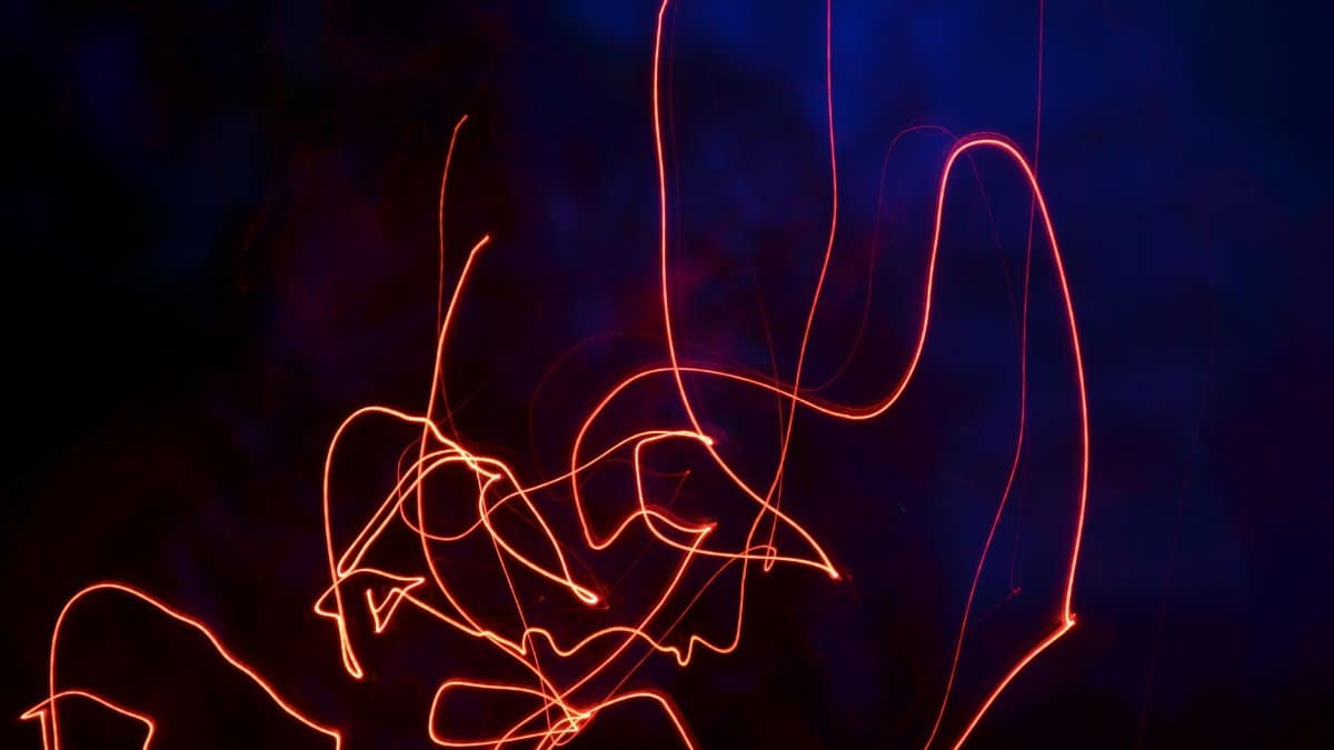
In laboratories around the nation researchers have found a way to follow the swift electrical impulses that pass between brain cells. This method called Neuron Communication Imaging captures those signals as they occur and opens a window onto the basic operations of the mind. The advance relies on a fluorescent marker that lights up in response to voltage changes and thereby reveals patterns once hidden from view. Such observations may help explain how networks of cells give rise to memory movement and perception itself.
Tracing Voltage Changes Across Cell Networks

The fluorescence approach rests on a dye that shifts its glow according to the electrical state inside a neuron. When an impulse travels along the cell membrane the marker brightens for a fraction of a second and cameras record the event at high speed. This process lets observers follow activity through entire circuits rather than single points.
Advantages Over Older Recording Methods

Traditional electrodes can measure voltage yet they often disturb nearby tissue and sample only a few locations at once. The optical technique avoids physical contact and gathers data from thousands of cells in the same moment. As a result maps of signal flow become far more complete and less prone to artifacts from insertion.
Applications in Studies of Learning and Memory

Scientists have begun to apply Neuron Communication Imaging to brain regions known for storing experience. Early trials show distinct waves of activity that appear when animals encounter new tasks and later repeat them. These patterns suggest that timing between cells plays a larger role in forming lasting connections than previously measured.
Links to Disorders Involving Signal Disruption
Conditions such as epilepsy and certain forms of dementia involve irregular firing among neurons. By watching communication in real time researchers can identify the precise moments when order breaks down. The resulting data may guide the design of interventions that restore balanced activity without broad side effects.
Technical Challenges in Marker Development

Creating a reliable fluorescent sensor requires balancing brightness speed and safety inside living tissue. Teams continue to refine the molecules so they respond within microseconds yet remain harmless over long sessions. Progress in chemistry has already extended observation windows from seconds to minutes in animal models.
Potential for Exploring Consciousness and Mind

Beyond clinical uses the method invites reflection on how coordinated electrical events give rise to awareness. Spiritual traditions have long described inner experience as a flow of subtle energies and modern imaging now renders those flows visible in concrete terms. Such convergence may encourage dialogue between empirical findings and longstanding questions about the nature of self.
Future Integration With Other Imaging Tools
Researchers plan to combine the voltage sensor with markers for chemical messengers and structural proteins. The combined view would show both the electrical message and the molecular aftermath in the same cells. This layered picture could clarify how brief impulses lead to lasting structural change.
Broader Impact on Brain Research Funding

The visibility of the technique has drawn support from agencies that seek practical returns on basic science. Projects now span multiple institutions and share standardized protocols for data collection. Wider adoption should accelerate discovery while reducing duplication of effort across laboratories.
Ethical Considerations in Human Applications

Any move toward people will require careful review of safety and consent procedures. Noninvasive optical methods may eventually allow monitoring during routine medical care yet questions remain about privacy when brain activity is recorded in detail. Guidelines developed now will shape responsible use for years ahead.